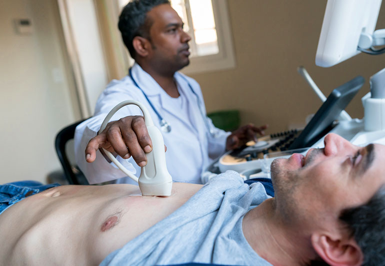
Heart conditions are increasing worldwide, including in Port Saint Lucie, Florida. An echocardiogram is a valuable diagnostic tool used by physicians to assess such heart conditions. It uses ultrasound waves to create an image of the heart, which helps diagnose conditions such as valvular heart disease, congenital heart defects, and cardiac tumors. Here is a list of six issues a Port Saint Lucie echocardiogram specialist can detect.
-
Valve Malfunctioning
Valves are an essential part of the heart as they maintain blood flow within the heart chambers. Various types exist, including those controlling blood from being pumped backward from one section to another. Another type includes blood moving from a ventricle into the atrium or from either ventricle back to its respective atrium.
Any problems with these valves could lead to what is called a valve prolapse. The images below show two examples of valvular malfunctions referred to as stenosis and insufficiency, respectively.
-
Blood Flow Through the Heart
The heart to receive oxygenated blood from the lungs uses a sizable pulmonary trunk vessel. In addition, another large vessel supplies blood to the body, known as the aorta. The echocardiogram can detect reduced or increased blood flow through these vessels, suggesting a blockage of the vessels. Critical stenosis of either vessel is considered a medical emergency as it can lead to heart failure or sudden death if not treated quickly.
-
Thickness and Size of the Chambers
The heart needs to be full of blood for the heart to pump correctly. A normal-sized shaped left ventricle will produce higher pressure to push enough blood into the aorta. Obstructed valves may lead to less than optimal filling (i.e., diastolic dysfunction), while diseases such as cardiomyopathy may lead to an enlarged heart. If any of these issues are present, the left ventricle will appear abnormally thick on the echocardiogram.
-
Blood Clots in the Heart
An echocardiogram can detect the presence of blood clots by using Doppler ultrasound. These clots may form in a chamber and cause valvular dysfunction or endocarditis (infection on the heart valves). The image below shows some poor valve function and also what appears to be one large clot that is restricting blood from being appropriately pumped and is probably the cause of the patient’s recent stroke (note: this is a lot easier to diagnose with an echocardiogram than it would be by looking at plain CT images).
-
Weak or Damaged Cardiac Muscle Tissue
Cardiomyopathy is a general term used to describe diseases that weaken or damage the heart’s normal muscle tissue. The LV chamber size and shape may be abnormal due to either dilation or hypertrophy, as seen in the images below. In addition, it detects other conditions such as cancer and inflammation of the heart muscle (myocarditis).
-
Pericardium Problems
The pericardium is a sac that surrounds the heart and even produces some of its fluids. When this sac becomes inflamed or forms scar tissue, it can interfere with the proper function of the heart by interfering with the diastolic filling phase of the cardiac cycle. The echocardiogram will pick up on this problem due to the abnormal structure.
An echocardiogram is a crucial diagnostic tool used by physicians to assess the condition of the heart. When using ultrasound waves, an image of the heart is created, which can diagnose conditions such as valvular heart disease, congenital heart defects, and cardiac tumors. In addition, various other issues with the heart can be detected, including blood clots, cardiomyopathy, and pericardium problems.


